Anatomy of the Bladder
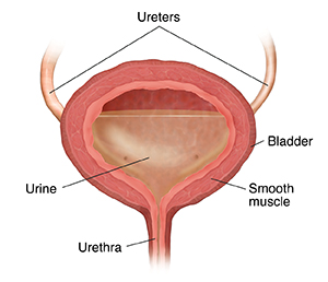
The bladder is part of your urinary tract. It's a hollow organ in your lower belly (pelvis). Urine is stored in it. This is the liquid waste that's made by the kidneys. Urine flows away from each kidney through a tube called a ureter. The ureters carry the urine into your bladder. The urine stays in your bladder until you let it pass out of your body through another tube called the urethra. You use ring-shaped muscles called sphincter muscles to control urine flow.
An outer layer of smooth muscle called the detrusor muscle surrounds the bladder. When your bladder is full, the muscles in the bladder wall can be tightened to squeeze out the urine. As you urinate, the bladder shrinks in size.
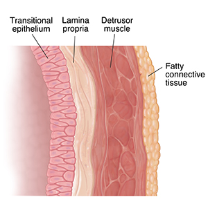
The bladder wall is made of many layers, including:
Urothelium or transitional epithelium. This is the layer of cells that lines the inside of the kidneys, ureters, bladder, and urethra. Cells in this layer are called urothelial cells or transitional cells.
Lamina propria. This is the next layer around the urothelium. It's a type of connective tissue.
Detrusor muscle (muscularis propria). This is the outer layer. It's the thick smooth muscle tissue outside the lamina propria.
Fatty connective tissue. This covers the outside of the bladder and separates it from other organs.
Superficial bladder cancer affects only the inside lining of the bladder. This is the transitional epithelium. Cancer here is called transitional cell carcinoma or urothelial carcinoma. As the cancer grows, it can become invasive bladder cancer that goes into deeper layers of the bladder wall. Over time, it can grow into the fatty connective tissue.

